Electron microscopy
Locations : UCBL Lyon 1, ENS de Lyon
Lab manager : Clémentine Fellah
Contact : clementine.fellah@ens-lyon.fr
Photo Vincent Moncorgé Ⓒ
Introducing the facility
The electron microscopy platform provides all the laboratory’s research themes with imaging techniques for observing and analyzing the surface of natural and experimental samples. It is possible to obtain small-scale information on the morphology, structural organization and elemental composition of a wide range of materials.
These facilities are available to LGL-TPE researchers, but are also open to any external scientific collaboration requiring expertise in materials characterization at the microscopic scale. For training, reservation or development requests, please contact the platform’s lab manager.
The platform’s instruments for sample preparation and analysis are spread over several sites, at the University Claude Bernard, UCBL Lyon 1 and Ecole normale supérieure de Lyon, ENS de Lyon.
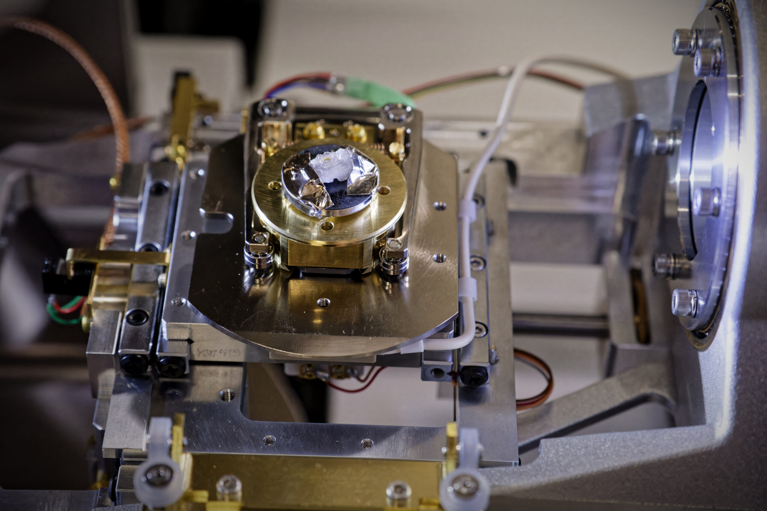
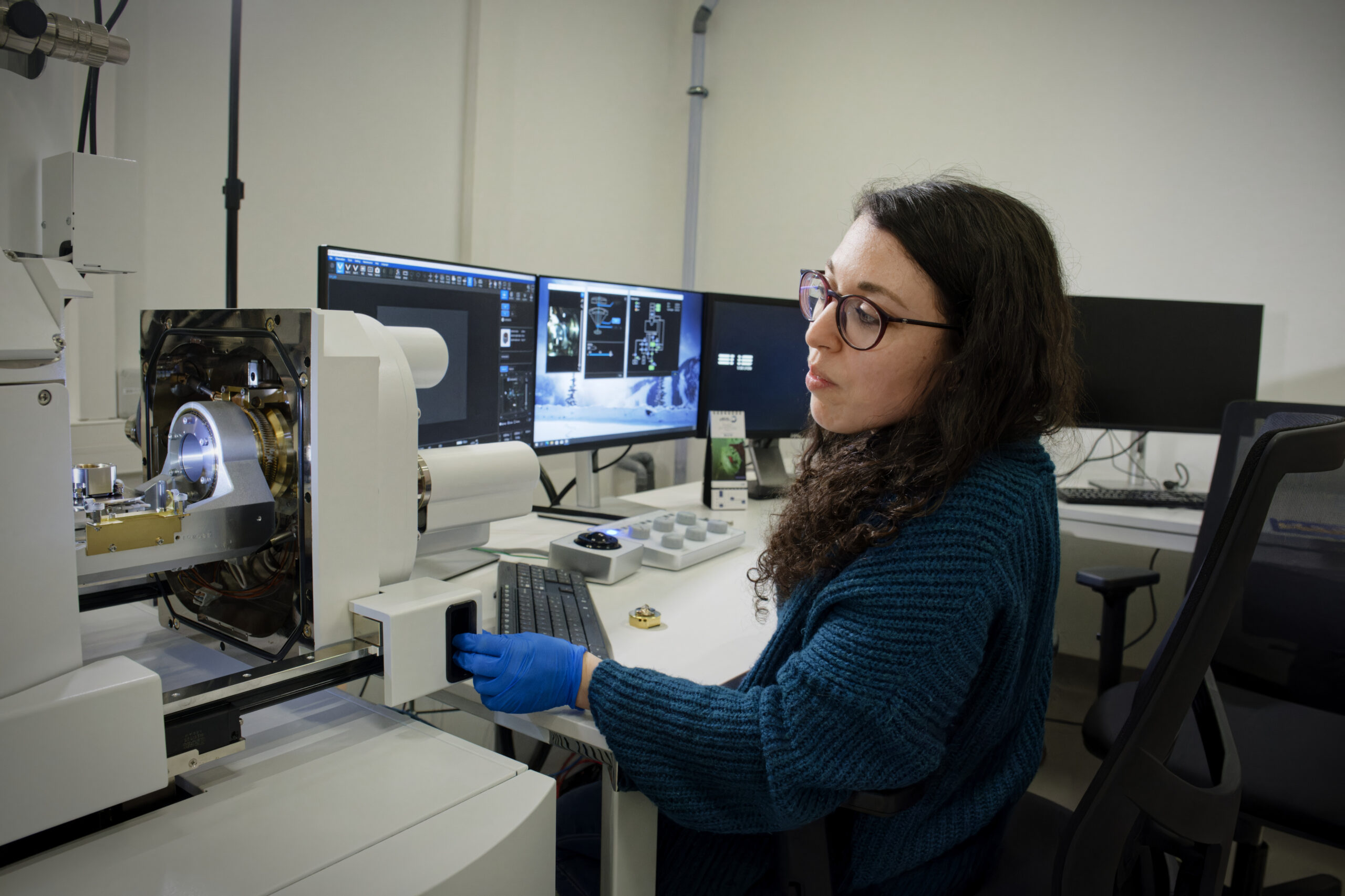
Sample preparation (UCBL campus – Géode building- and ENS de Lyon campus – M8 building)
Several cutting, coating and polishing tools are available at both Lyon campus to prepare bulk samples for electron microscopy and micro-analysis. A LEICA EM ACE600 carbon evaporator is also available at the ENS de Lyon for the preparation of low-conductivity or insulating samples. Photo Vincent Moncorgé Ⓒ
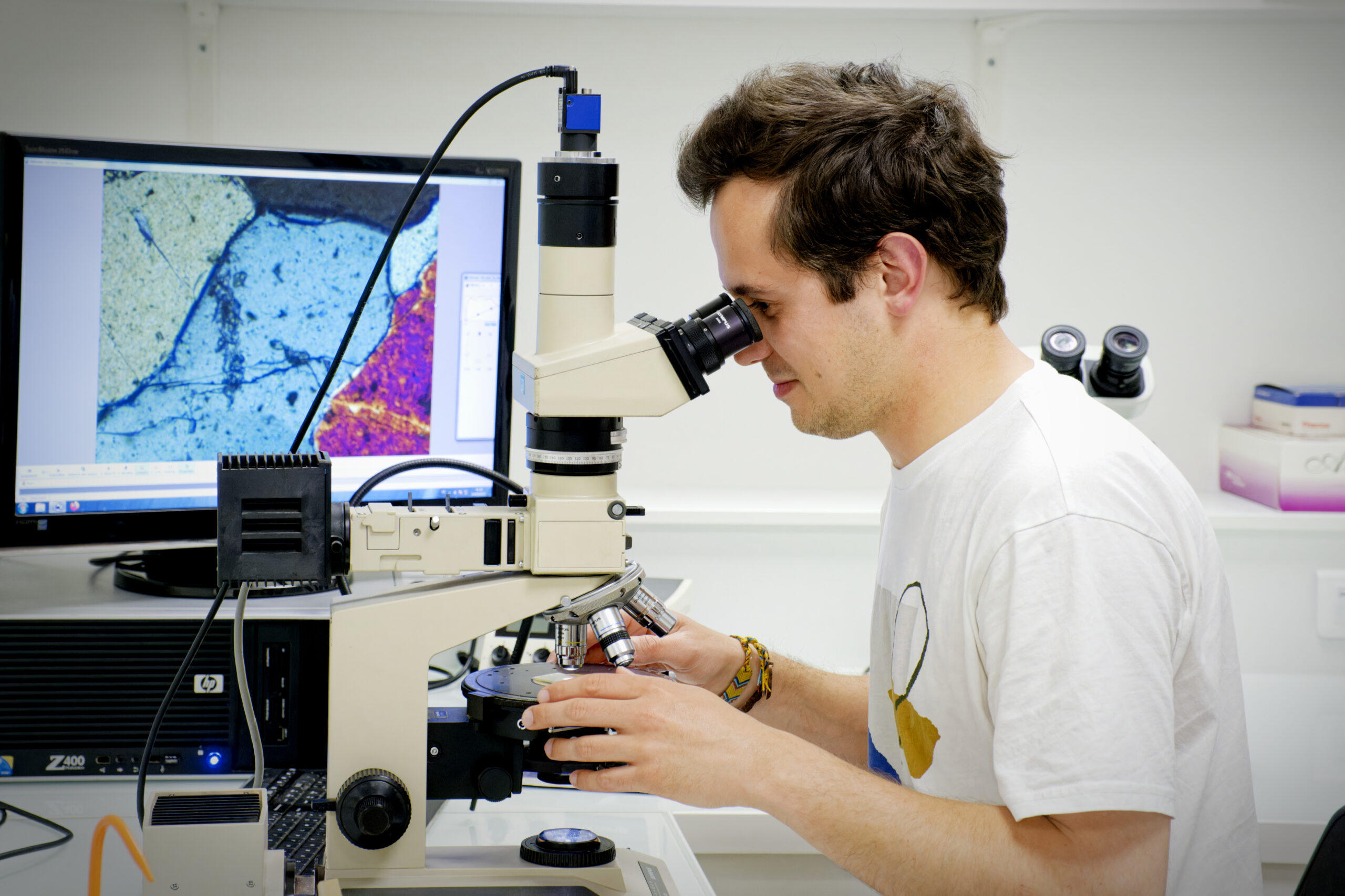
Observations and analysis (ENS de Lyon)
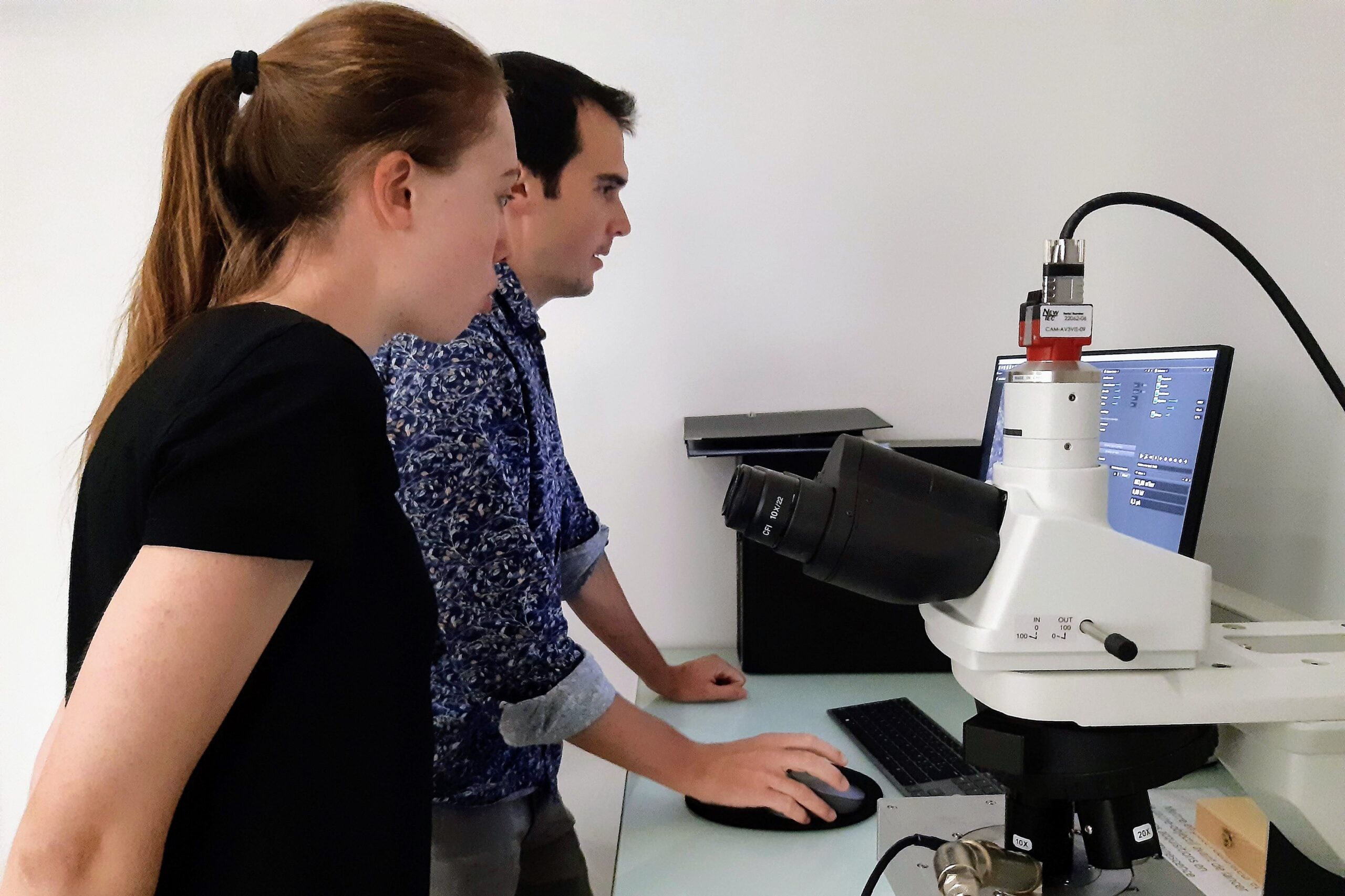
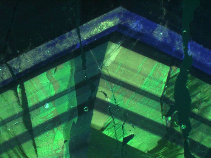
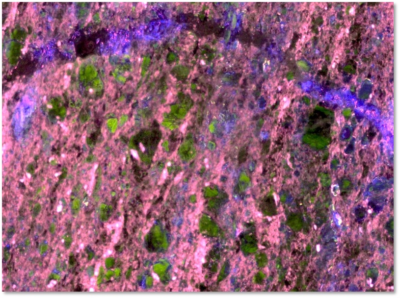
Cathodoluminescence microscope
The Cathodyne cathodoluminescence facility (NewTec Scientific) can image very fine variations in trace element concentration from the centimeter to the micrometer scale. Sample structures (zonation, oscillations, grains) can easily be detected on the surface before moving on to smaller-scale characterization on the SEM. Photo Vincent Moncorgé Ⓒ
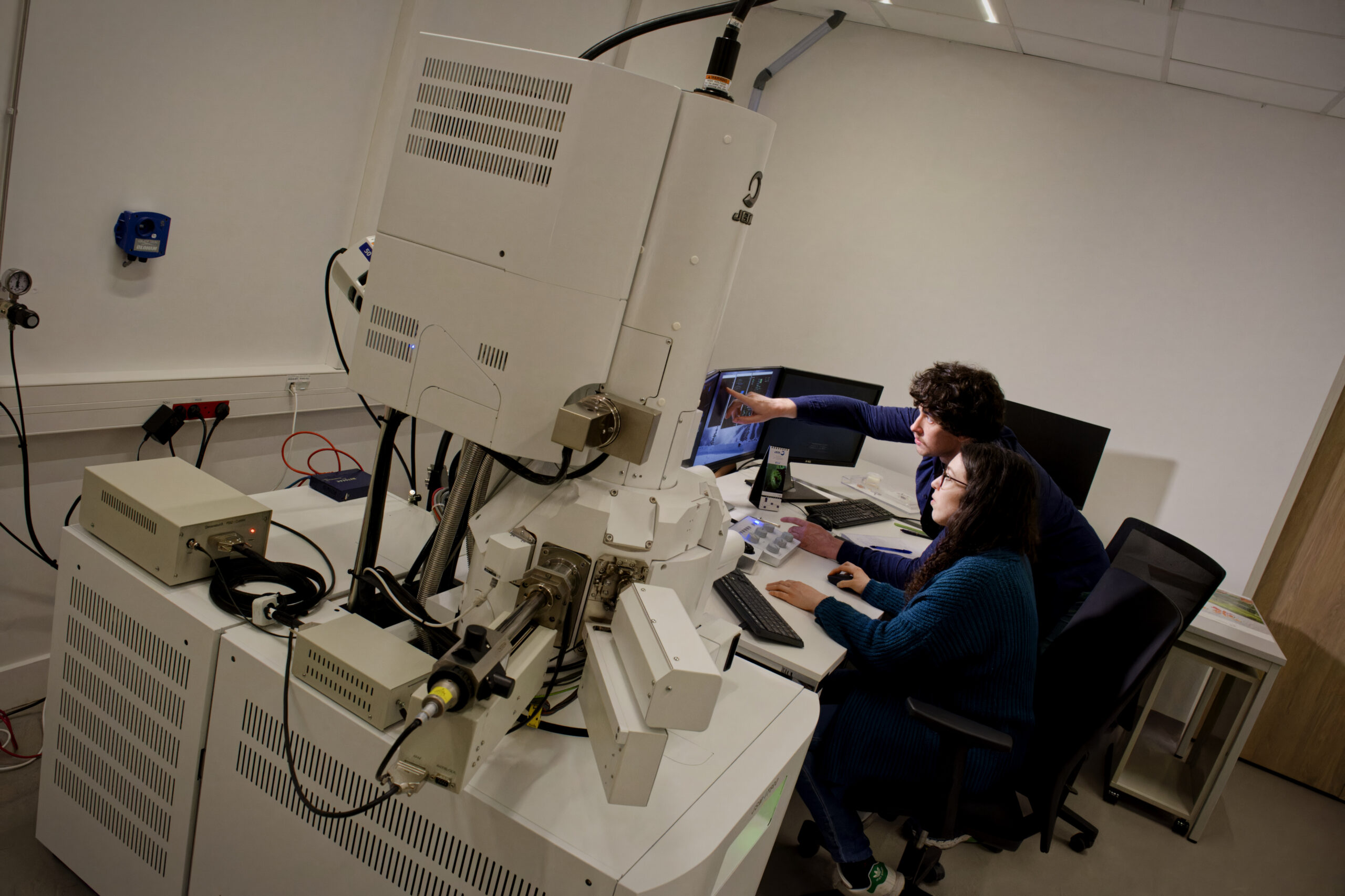
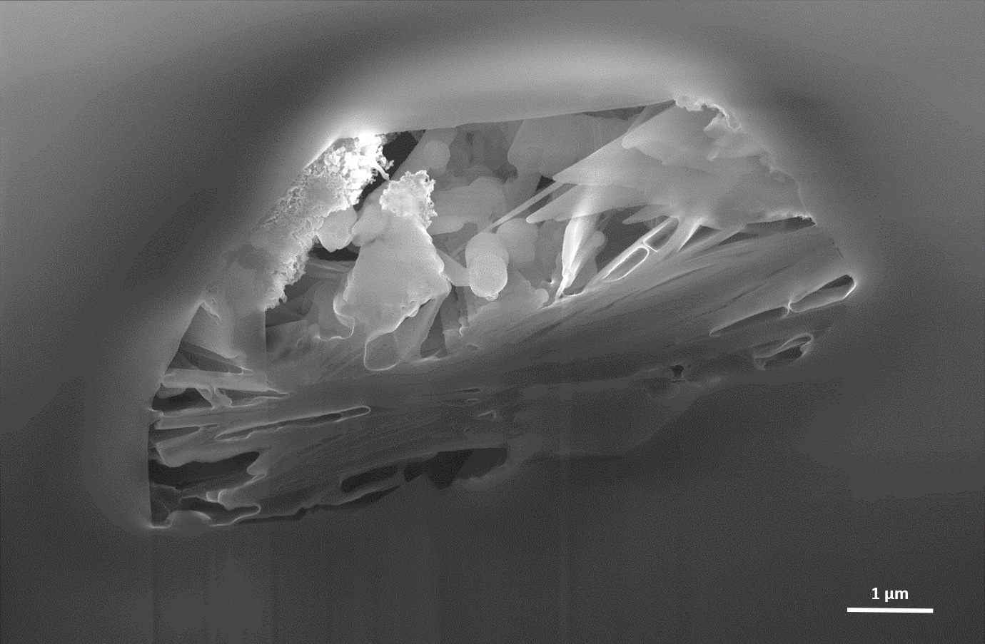
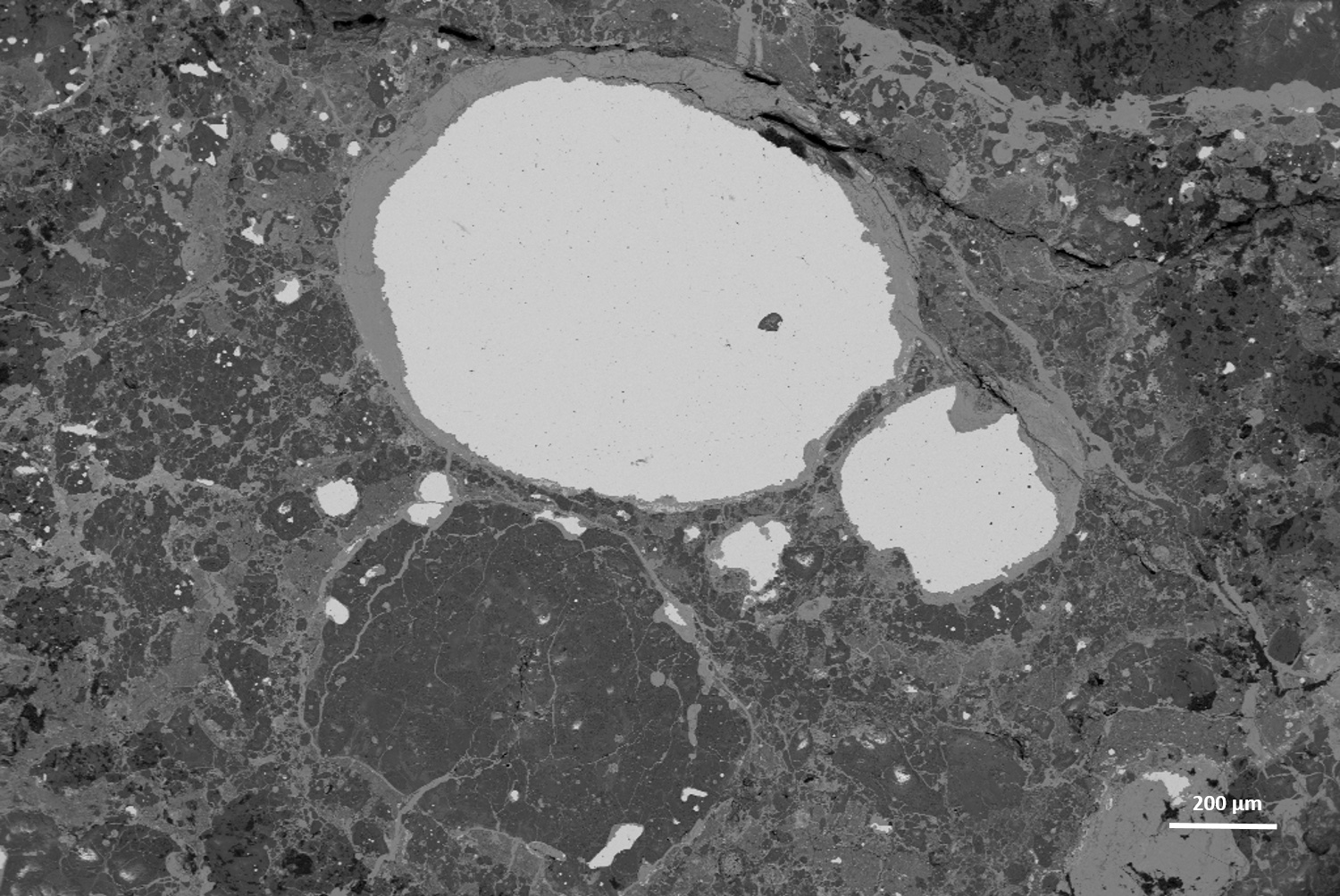
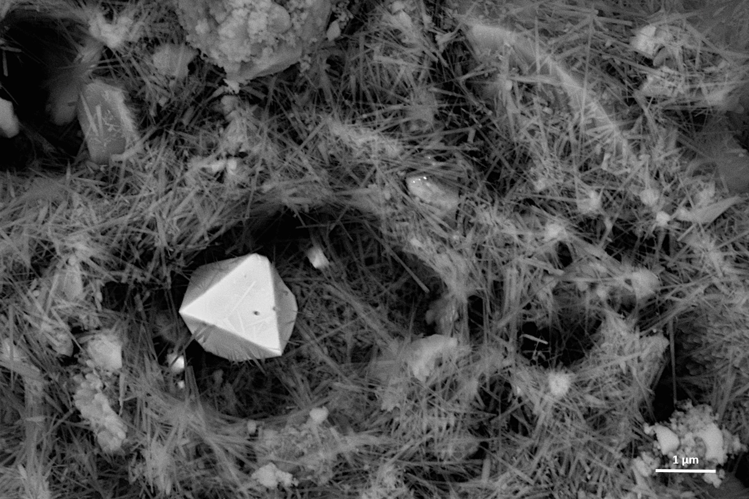
Scanning electron microscope (SEM)
The SEM is a technique for high-resolution imaging and analysis of sample surfaces. The platform’s IT800HL (JEOL) facility is equipped with imaging detectors, a HAADF SEM STEM detector for thin samples and an EDX detector (X-Max 50 from Oxford Instruments) for elemental micro-analysis. This SEM can analyze nanoscale samples in partial vacuum up to 300 Pa. Photo Vincent Moncorgé Ⓒ
The LGL – TPE is a partner of CLYM (Consortium Lyon St-Etienne de Microscopie – FED 4092) and has privileged access to state-of-the-art electron microscopy facility at the INSA Lyon (https://www.clym.fr/fr/microscopes; Contact: C. Fellah), at the CNRS (https://www.clym.fr/fr/node/71), and at the University Jean Monnet of Saint-Etienne (last generation of corrected TEM NeoARM200F: https://www.clym.fr/fr/node/336 et https://www.clym.fr/fr/node/441 – Contact: A-M Seydoux-Guillaume).